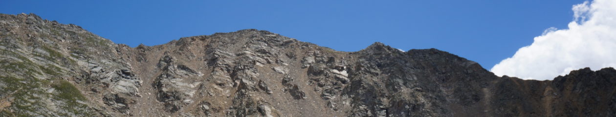
Can I Ever Go Back Up To High Altitude Again? – Recurrence Risk of HAPE & HARPE

by Taylor Kligerman, PA-S

Can I ever return to high altitude? Do I need to move down to a lower elevation?

Disease processes often differ at high altitudes. Some conditions have only been known to occur at high elevations. Most of the resources cited in this blog refer to ‘high altitude’ being at or above 2,500 meters or 8,200 feet.

Ebert Family Clinic in Frisco, Colorado is at 9,075 ft. Many areas in the immediate vicinity are over 10,000′, with some patients living above 11,000′. Two of the more common conditions seen in patients at Ebert Family Clinic are high altitude pulmonary edema (HAPE) and high altitude resident pulmonary edema (HARPE), similar conditions that affect slightly different populations in this region of the Colorado Rocky Mountains.

In “classic” HAPE, a visitor may come from a low-altitude area to Frisco on a trip to ski with friends. On the first or second day, the person notices a nagging cough. They might wonder if they caught a virus on the plane ride to Denver. The cough is usually followed by shortness of breath that begins to make daily tasks overwhelmingly difficult. One of the dangerous aspects of HAPE is a gradual onset leading patients to believe their symptoms are caused by something else. A similar phenomenon is seen in re-entry HAPE, where a resident of a high altitude location travels to low altitude for a trip and upon return experiences these same symptoms [1].

In HARPE, a person living and working here in Frisco may be getting ill or slowly recovering from a viral illness and notices a worsening cough and fatigue. These cases are even more insidious, going unrecognized, and so treatment is sought very late. Dr. Christine Ebert-Santos and her team at Ebert Family Clinic hypothesize that while residents have adequately acclimated to the high-altitude environment, the additional lowering of blood oxygen due to a respiratory illness with inflammation may be the inciting event in these cases.

In both cases, symptoms are difficult to confidently identify as a serious illness versus an upper respiratory infection, or simply difficulty adjusting to altitude. For this reason, Dr. Chris recommends that everyone staying overnight at high altitude obtain a pulse oximeter. Many people became familiar with the use of these instruments during the COVID-19 pandemic. The pulse oximeter measures what percent of your blood is carrying oxygen. At high altitude, a healthy level of oxygenation is typically ≥90%. This is an easy way to both identify potential HAPE/HARPE, as well as reassure patients they are safely coping with the high-altitude environment [2].

HAPE and HARPE are both a direct result of hypobaric hypoxia, a lack of oxygen availability at altitude due to decreased atmospheric pressures. At certain levels of hypoxia, we observe a breakdown in the walls between blood vessels and the structures in lungs responsible for oxygenating blood. The process is still not totally understood, but some causes of this breakdown include an inadequate increase in breathing rates, reduced blood delivered to the lungs, reduced fluid being cleared from the lungs, and excessive constriction of blood vessels throughout the body. These processes cause fluid accumulation throughout the lungs in the areas responsible for gas exchange making it harder to oxygenate the blood [3].

We do know that genetics play a significant role in a person’s risk of developing HAPE/HARPE. Studies have proposed many different genes that may contribute, but research has not, so far, given healthcare providers a clear picture of which patients are most at-risk. Studies have shown that those at higher risk of pulmonary hypertension (high blood pressure in the blood vessels of your lungs), are more likely to develop HAPE [4]. This includes some types of congenital heart defects [5,6]. High blood pressures in the lungs reach a tipping point and appear to be the first event in this process. However, while elevated blood pressures in the lungs are essential for HAPE/HARPE, this by itself, does not cause the condition. The other ingredient necessary for HAPE/HARPE to develop is uneven tightening of the blood vessels in the lungs. When blood vessels are constricted locally, the blood flow is shifted mainly to the more open vessels, and this is where we primarily see fluid leakage. As the blood-oxygen barrier is broken down in these areas, we may also see hemorrhage in the air sacs of the lungs [3].

One observation healthcare providers and scientists have observed is that HAPE/HARPE can be rapidly reversed by either descending from altitude or using supplemental oxygen. Both strategies increase the availability of oxygen in the lungs, reducing the pressure on the lungs’ blood vessels by vasodilation, quickly improving the integrity of the blood-oxygen barrier.

In a preliminary review of over 100 cases of emergency room patients in Frisco diagnosed with hypoxemia (low blood oxygen content) Dr. Chris and her team have begun to see trends that suggest the availability of at-home oxygen markedly reduces the risk of a trip to the hospital. This demonstrates that patients with both at-home pulse oximeters and supplemental oxygen have the capability to notice possible symptoms of HAPE, assess their blood oxygen content, and apply supplemental oxygen if needed. This stops the development of HAPE/HARPE before damage is done in the lungs. In the case of many of our patients, these at-home supplies prevent emergencies and allow patients time to schedule an appointment with their primary care provider to better evaluate symptoms.

Additionally, Dr. Chris and her team have observed that patients with histories of asthma, cancer, pneumonia, and previous HAPE/HARPE are often better educated and alert to these early signs of hypoxia and begin treatment earlier on in the course of HAPE/HARPE, reducing the relative incidence identified by medical facilities. There are many reasons to seek emergent care such as low oxygen with a fever. Patients with other existing diseases causing chronically low oxygen such as chronic lung disease may not be appropriately treated with  supplemental oxygen, although this is a very small portion of the population. Discussions with healthcare providers on the appropriate prevention plan for each patient will help educate and prevent emergency care visits in both residents and visitors.

A young child with short brown hair and glasses with dark, round frames wears a nasal canula for oxygen.

Studies of larger populations have yet to be published. A review of the case reports in smaller populations suggests that the previously estimated recurrence rate of 60-80% is exaggerated. This is a significant finding as healthcare providers have relied on this recurrence rate to make recommendations to their patients who have been diagnosed with HAPE. A review of 21 cases of children in Colorado diagnosed with HAPE reported that 42% experienced at least one recurrence [7]. This study was conducted by voluntary completion of a survey by the patients (or their families) which could lead to significant participation bias affecting the results. Patients more impacted by HAPE are more likely to complete these surveys. Another study looking at three cases of gradual re-ascent following an uncomplicated HAPE diagnosis showed no evidence of recurrence. The paper also suggested there may be some remodeling of the lung anatomy after an episode of HAPE that helps protect a patient from reoccurrence [8]. Similar suggestions of remodeling have been proposed through evidence of altitude being a protective factor in preventing death as demonstrated by fatality reports from COVID-19[9].

Without larger studies and selection of participants to eliminate other variables like preexisting diseases, we are left to speculate on the true rate of reoccurrence based on the limited information we have. Strategies to reduce the risk of HAPE/HARPE such as access to supplemental oxygen, pulse oximeters, and prescription medications [10] are the best way to prevent HAPE/HARPE. Research should also continue to seek evidence of individuals most at risk for developing HAPE/HARPE [11].

A woman with reddish-brown, straight hair just below her shoulders, wears a white coat over a mustard-colored shirt, smiling.
  1. Ucrós S, Aparicio C, Castro-Rodriguez JA, Ivy D. High altitude pulmonary edema in children: A systematic review. Pediatr Pulmonol. 2023;58(4):1059-1067. doi:10.1002/ppul.26294
  2. Deweber K, Scorza K. Return to activity at altitude after high-altitude illness. Sports Health. 2010;2(4):291-300. doi:10.1177/1941738110373065
  3. Bärtsch P. High altitude pulmonary edema. Med Sci Sports Exerc. 1999;31(1 Suppl):S23-S27. doi:10.1097/00005768-199901001-00004
  4. Eichstaedt C, Benjamin N, Grünig E. Genetics of pulmonary hypertension and high-altitude pulmonary edema. J Appl Physiol. 2020;128:1432
  5. Das BB, Wolfe RR, Chan K, Larsen GL, Reeves JT, Ivy D. High-Altitude Pulmonary Edema in Children with Underlying Cardiopulmonary Disorders and Pulmonary Hypertension Living at Altitude. Arch Pediatr Adolesc Med. 2004;158(12):1170–1176. doi:10.1001/archpedi.158.12.1170
  6. Liptzin DR, Abman SH, Giesenhagen A, Ivy DD. An Approach to Children with Pulmonary Edema at High Altitude. High Alt Med Biol. 2018;19(1):91-98. doi:10.1089/ham.2017.0096
  7. Kelly TD, Meier M, Weinman JP, Ivy D, Brinton JT, Liptzin DR. High-Altitude Pulmonary Edema in Colorado Children: A Cross-Sectional Survey and Retrospective Review. High Alt Med Biol. 2022;23(2):119-124. doi:10.1089/ham.2021.0121
  8. Litch JA, Bishop RA. Reascent following resolution of high altitude pulmonary edema (HAPE). High Alt Med Biol. 2001;2(1):53-55. doi:10.1089/152702901750067927
  9. Gerken J, Zapata D, Kuivinen D, Zapata I. Comorbidities, sociodemographic factors, and determinants of health on COVID-19 fatalities in the United States. Front Public Health. 2022;10:993662. Published 2022 Nov 3. doi:10.3389/fpubh.2022.993662
  10. Luks A, Swenson E, Bärtsch P. Acute high-altitude sickness. European Respiratory Review. 2017;26: 160096; DOI: 10.1183/16000617.0096-2016
  11. Dehnert C, Grünig E, Mereles D, von Lennep N, Bärtsch P. Identification of individuals susceptible to high-altitude pulmonary oedema at low altitude. European Respiratory Journal 2005;25(3):545-551; DOI: 10.1183/09031936.05.00070404

Hypoxia in the Emergency Department: Preliminary Analysis of Data from the Highest Atitude Population in North America & Children with Hypoxia

Hypoxia is a common presentation at the emergency department for the St Anthony Summit Medical Center, located at 2800 meters above sea level (msl) in Colorado. Children under 18 are brought in with respiratory symptoms, trauma, congenital heart and lung abnormalities, and high altitude pulmonary edema (HAPE). Many complain of shortness of breath and/or cough and are found to be hypoxic, defined as an oxygen saturation below 89% on room air for this elevation. Patients who live at altitude may perform home pulse oximetry and arrive for treatment and diagnosis of known hypoxia. Extensive and ongoing analysis of the data from children found to be hypoxic in the emergency department raises many questions, including how residents vs nonresidents present, how often  these cases are preceded by febrile illness and what chief complaint is most frequently cited. 

Understanding the presentation of hypoxia in children at altitude can help ensure that healthcare providers are following a comprehensive approach with awareness of the overlapping symptoms of HAPE, pneumonia and asthma. Below is a graphic summary of 36 cases illustrating the clinical, social and geographic factors contributing to hypoxia at altitude in residents and visitors. A further analysis of over 200 children with hypoxia presenting to the emergency room at 9000 feet is underway including x-ray findings.

The graphs below were created by the author, using data extracted directly from a review of patient charts (specifically, those of children presenting to the local hospital in Summit County, Colorado (9000 feet) with hypoxia).

Graphs 1-4 show chief complaints of cough (CC) and shortness of breath (SOB) compared by age and by residence (residence includes altitudes above 2100 msl, the front range (a high altitude region of the Rocky Mountains running north-south between Casper, Wyoming and Pueblo, Colorado) averaging 1500 msl, and out of the state of Colorado) 

A graph of age with blue bars

Description automatically generated
A graph with blue bars

Description automatically generated
A graph with blue bars

Description automatically generated
A graph of a bar chart

Description automatically generated with medium confidence

Graphs 5-6 show presence of fever by residence and by age 

A graph with blue bars

Description automatically generated
A graph of a number of people with fever

Description automatically generated

Graphs 7-8 show presence of asthma by residence and by age 

A graph with blue bars

Description automatically generated
A graph with blue bars

Description automatically generated

Graphs 9 and 10 show lowest oxygen by age at admission and lowest O2 organized by days spent in the county (residents are excluded from this data). 

A graph with blue dots

Description automatically generated
A graph with blue dots

Description automatically generated

Doc Talk: Physician Altitude Experts on High Altitude Pulmonary Edema (HAPE)

One of our students recently came across a comprehensive publication on high altitude pulmonary edema (HAPE) on reputable point-of-care clinical resource UpToDate.com1, citing Christine Ebert-Santos, MD, MPS, the founder of highaltitudehealth.com.

Emergency medicine physician at Aspen Valley Hospital and medical director for Mountain Rescue Aspen since 1997 Dr. Scott A. Gallagher2 and emergency physician and altitude research pioneer Dr. Peter Hackett3 introduce the resource warning, “Anyone who travels to high altitude, whether a recreational hiker, skier, mountain climber, soldier, or worker, is at risk of developing high-altitude illness.”

Ebert-Santos’s (known affectionately to her patients and mountain community as “Dr. Chris”) own research is referenced in the article’s discussion of epidemiology and risk factors noting an additional category of HAPE among “children living at altitude who develop pulmonary edema with respiratory infection but without change in altitude,”4 whereas the two other recognized categories (classic HAPE and re-entry HAPE) typically happen in response to a change in altitude.

The article continues with figures illustrating how ascending too quickly or too much can dramatically increase risk: “HAPE generally occurs above 2500 meters (8000 feet) and is uncommon below 3000 meters (10,000 feet) … The risk depends upon individual susceptibility, altitude attained, rate of ascent, and time spent at high altitude. in those without a history of HAPE, the incidence is 0.2 percent with ascent to 4500 meters (14,800 feet) over four days but 6 percept when ascent occurs over one to two days. In those with a history of HAPE, recurrence is 60 percent with an ascent to 4500 meters over two days. At 5500 meters (18,000 feet), the incidence ranges between 2 and 15 percent, again depending upon rate of ascent.”

Dr. Chris discusses her experience treating her pediatric patients at high altitude in more depth in an interview with pediatric emergency medicine physician Dr. Alison Brent from Colorado Children’s Hospital for the podcast Charting Pediatrics.

Dr. Gallagher and Dr. Hackett’s article is available on UpToDate with a subscription.

  1. https://www.uptodate.com/contents/high-altitude-pulmonary-edema?source=autocomplete&index=0~1&search=HAPE ↩︎
  2. https://www.aspenhospital.org/people/scott-a-gallagher-md/ ↩︎
  3. https://www.highaltitudedoctor.org/dr-peter-hackett ↩︎
  4. Ebert-Santos, C. High-Altitude Pulmonary Edema in Mountain Community Residents. High Alt Med Biol 2017; 18:278. ↩︎

Interview with Dr. Christine Ebert-Santos on High Altitude Pulmonary Edema

by Cody Jones, Summit Daily News

“‘The first sign is usually a cough,’ Ebert-Santos said. ‘Followed by shortness of breath with any effort — even just walking — and fatigue. You just want to lie on the couch.’

If left untreated the early warning signs of high altitude pulmonary edema can rapidly progress into having fluid build up in the lungs, which will then lead to a patient’s oxygen saturation levels rapidly decreasing. If the individual does not seek treatment quickly, the condition can be fatal.”

Read the whole article here.

The Impact of High Altitude on Diabetes Diagnosis: The Relationship between Hemoglobin A1c and Fasting Plasma Glucose

Type 2 Diabetes (T2D) has emerged as a global concern, with its prevalence steadily increasing. The test of choice to diagnose and monitor T2D is hemoglobin A1c (HbA1c), which tracks average blood sugar levels over the last three months. Normal HbA1c levels are below 5.7%, 5.7% to 6.4% indicates prediabetes, and 6.5% or higher indicates diabetes. Within the prediabetes range, high HbA1c levels increase the risk of developing T2D. Additionally, levels above 6.5% correlate with greater risk for diabetes complications.1 Fasting Plasma Glucose (FPG) is an additional test that indicates an immediate blood sugar level following a period of fasting. Normal FPG levels are below 100 mg/dL (5.5 mmol/L), 100 to 125 mg/dL (5.6 to 6.9 mmol/L) suggests prediabetes, whereas 126 mg/dL (7 mmol/L) or higher generally indicates diabetes.2 Because HbA1c provides an overview of blood sugar levels spanning the past 2-3 months, it offers a more comprehensive insight into blood sugar management and is the preferred diagnostic test for T2D.3 Recent studies are unveiling discrepancies between HbA1c and glucose testing, prompting discussions on specific diagnostic criteria for different populations.

People living at high altitude experience unique physiological adaptations, such as higher hemoglobin levels and specific glucose metabolism patterns. Acknowledging these adaptations, a 2017 study by Bazo-Alvarez et. al sought to evaluate the relationship between HbA1c and FPG among individuals at sea level compared to those at high altitude.

The study analyzed data from 3613 Peruvian adults without diagnosed diabetes from both sea level and high altitude (>3000m). The mean values for hemoglobin, HbA1c, and FPG differed significantly between these populations. The correlation between HbA1c and FPG was quadratic at sea level but linear at high altitude, suggesting different glucose metabolism patterns. Additionally, for an HbA1c value of 48 mmol/mol (6.5%), corresponding mean FPG values were significantly different: 6.6 mmol/l at sea level versus 14.8 mmol/l at high altitude.

Tall, snowy mountain peaks rise in the distance over rows of deep green pine trees growing out of the hills around a bike. path in the foreground.

This significant difference in predictive values suggests potential controversy in utilizing HbA1c as a diagnostic tool for diabetes in high altitude settings. Using HbA1c at altitude potentially underdiagnoses and under treats patients. To ensure a more accurate diagnosis of T2D at high altitude, reevaluating diagnostic criteria, possibly leaning towards FPG or oral glucose tolerance testing (OGTT) might be necessary.

In conclusion, this study emphasizes the need for careful consideration when diagnosing diabetes in high-altitude regions. Future research is warranted, including studies replicating the findings of the cross-sectional study by Bazo-Alvarez and longitudinal studies exposing the long-term effects of the diagnostic discrepancy of HbA1c in high altitude patients. This additional data will ensure accurate diagnosis and appropriate management of diabetic patients at high altitude.

  1. Centers for Disease Control and Prevention. A1C Test. Accessed 12/26/23. Available from: https://www.cdc.gov/diabetes/managing/managing-blood-sugar/a1c.html
  2. World Health Organization. Fasting Blood Glucose. Accessed 12/26/23. Available from: https://www.who.int/data/gho/indicator-metadata-registry/imr-details/2380#:~:text=When%20fasting%20blood%20glucose%20is,separate%20tests%2C%20diabetes%20is%20diagnosed   
  3. Sherwani, S.I., et al. 2016. Significance of HbA1c Test in Diagnosis and Prognosis of Diabetic Patients. Biomark. Insights. 2016 Jul; 11: 95-104. DOI: 10.4137/BMI.S38440.
  4. Bazo-Alvarez, J. C., et al. Glycated haemoglobin (HbA1c) and fasting plasma glucose relationships in sea-level and high-altitude settings. Diabet. Med. 2017 Jun; 34(6): 804-812. DOI: 10.1111/dme.13335.

What is Acute Mountain Sickness?

Acute mountain sickness (AMS) is a condition that can occur when individuals ascend to high altitudes rapidly, typically above 2,500 meters (8,200 feet). The symptoms of AMS are due to the body’s struggle to adapt to the decreased oxygen levels at higher elevations. More specifically, the symptoms are caused by cerebral vasodilation that occurs in response to hypoxia, in an attempt to maintain cerebral perfusion.1

The typical symptoms of AMS include headache, nausea, vomiting, anorexia, and fatigue. In children the symptoms are less specific including increased fussiness, crying, poor feeding, disrupted sleep, and vomiting. Symptom onset is usually 6-12 hours after arrival to altitude but this can vary.

AMS affects children, adults, males and females equally, with a slight increased incidence in females. It is difficult to believe, but physical fitness does not offer protection against AMS. However, people who are obese, live at low elevation, or undergo intense activities upon arrival to elevation are at increased risk.1

Descending

Descending and decreasing altitude is a vital treatment for people with severe symptoms of AMS. By decreasing altitude there will be more oxygen in the air and symptoms will not be as severe..2 

Oxygen

Since the main cause of AMS is hypoxia, oxygen supplementation is an effective treatment when descent is not wanted or possible. Supplemental oxygen even at .5L to 1L per hour can be effective in reducing symptoms.It can be prescribed for short periods of time or to be used only during sleep  In the central Colorado Rockies, this may be a practical solution for “out of towners” who have traveled up to the town of Leadville (10,158’/3096m) for vacation, but in an austere environment supplemental oxygen may not be a reasonable treatment option. There should be symptomatic improvement within one hour.

Acetazolamide

Acetazolamide is a carbonic anhydrase inhibitor which causes increased secretion of sodium, potassium, bicarb, and water. This mechanism of actions lends beneficial to the treatment of AMS because it decreases the carbonic anhydrase in the brain. 3There is evidence to support the use of acetazolamide in the prevention of AMS, but minimal evidence pointing towards it’s role in treatment. Dosing is inconsistent but is usually prescribed at 125-250mg BID.

Hyperbaric Therapy

Many people consider hyperbaric chambers to be large structures in hospitals, however there are portable and lightweight hyperbaric chambers that can be used in austere environments or during expeditions. The mechanism of action of hyperbaric therapy is a simulated decrease in elevation, of approximately 2500 meters. These chambers will remove symptoms within approximately one hour of use but symptoms are likely to return. They are useful in the field but not frequently required in a hospital setting.1

  1. https://www.uptodate.com/contents/acute-mountain-sickness-and-high-altitude-cerebral-edema?search=acute%20mountain%20sickness&source=search_result&selectedTitle=1~15&usage_type=default&display_rank=1#H35
  2. https://my.clevelandclinic.org/health/diseases/15111-altitude-sickness
  3. https://www.uptodate.com/contents/acetazolamide-drug-information?search=acetazolamide%20altitude&source=search_result&selectedTitle=2~150&usage_type=default&display_rank=2#F129759

High Altitude Sleep Disorders … A Thing of the Past?

The fundamentals of vitality include food, water, air, shelter, and sleep. Sleep, though often underappreciated, can influence our physical and mental  health,  complex and easily impacted by outside factors. Living at a  high altitude may be wonderful but what is gained in beauty and adventure, is compromised with  reduced quality sleep. With increasing elevation comes more nighttime awakenings,  brief arousals, nocturnal hypoxemia, and periodic breathing. Light  sleep increases and slow-wave and REM sleep decrease.

The current gold standard for diagnosis of suspected sleep disorders includes polysomnography:  seven or more streams of data at a hospital or sleep center. The SleepImage  System allows for more flexibility with children, adolescents, and adults. Currently,  Dr. Chris Ebert-Santos of Ebert Family Clinic in Frisco, Colorado, USA (9000′) is using this technology primarily to assess some of the most common  forms of Sleep Breathing Disorders and secondly, to analyze the percentage of oxygen  desaturation of her patients while in their homes. 

The SleepImage System measures several variables that construct a summary for each  individual. Sleep quality is generated using Sleep Quality Index (SQI) biomarkers. Pathology  markers measure sleep duration, efficiency, and latency. Central Sleep Apnea (CSA) and Obstructive Sleep Apnea (OSA) are assessed together as Sleep Apnea Hypoxia Index (sAHI). Periodic and fragmented sleep pathology are reported and can be used to assess disease  management long-term. 

Recently, the clinic analyzed Patient X’s sleeping patterns without and with  supplemental oxygen. The theory: adding a steady flow of oxygen to the  nightly sleep regimen reduced the total amount of time desaturating and severity of sleep  breathing disorders. On the night preceding treatment, Patient X experienced an SQI of 17  (expected >55) and efficiency at 95% (expected >85%) for overall sleep quality. Sleep  opportunity demonstrated a 0h:02m latency (expected <30m), and duration of 5h:47m (expected  7-9h); sAHI was marked as severe for both 4% and 3% desaturation with values at 34 and 61,  respectively (severe= >30.0 in adults). Fragmented sleep was at 55% (expected <15%) and  periodicity at 22% (expected <2%). Lastly, Patient X spent 25% of his night’s sleep under 90%  SpO2, 18% under 88% Spo2, and 4% under 80% SpO2. Ideally, a healthy night’s sleep should  aim to remain above 90% SpO2 for the majority of the time in bed. 

When oxygen supplementation was introduced, improvements were observed. Sleep quality  showed a slight change, SQI increased to 31 (previously 17, expected >55), and efficiency  decreased to 87% (previously 95%; expected >85%) while remaining at a target value. Sleep  opportunity showed a slight increase during latency to 0h:12m while remaining within the  expected value of <30mins; duration jumped to 8h:14m but that could be attributed to an early  bedtime. Fragmented sleep remained in the severe range but decreased by 5%; periodicity improved to 0%, removing it from both the severe and moderate range. The most notable  improvement was observed with sAHI, both the 3% and 4% desaturation categories improved to the moderate range with values of 9 and 14, respectively. Time under 90% SpO2 also improved  to only 4% throughout the night and 0% below 88% SpO2. 

Since data is collected while patients sleep, skewed results from the placebo effect can be  reduced or eliminated. Increased duration could be attributed to longer time in bed, as mentioned  above, and should be examined more in-depth longitudinally. Latency for sleep increased with  oxygen treatment but that could be attributed to discomfort from the nasal cannula or greater  tiredness one day over the other. Similarly, latency should be examined longitudinally.

The results seen with this patient are common in our population.  Many people report they slept significantly better their first night on oxygen. Many patients studied on and off oxygen show the same dramatic decrease in their sleep apnea index. The gold standard for treating sleep apnea involves a mask to increase the pressure in the airway and prevent the collapse and narrowing that occurs during relaxation and sleep.  Does the supplemental 2 liters per minute of oxygen cause enough increased airway pressure to prevent airway narrowing? Supplemental oxygen would not be considered for an intervention or treatment in other locations where sleep studies are conducted because they are not usually showing significant hypoxia. Does the improvement in oxygen, even if it is the difference between oxygen saturations in the high 80’s and low 90’s increasing to the mid 90’s affect the balance of oxygen and carbon dioxide in a way that changes the incidence of apnea and drive to breathe during sleep?

Long-term, this easy-to-use SleepImage System can assess sleep disorders  across all age groups and contribute to long-term management for many people living at altitude. Oxygen, a simple intervention that is widely available and relatively inexpensive, requiring no special visits to fit and adjust, has the potential to  improve symptoms and sleep greatly. 

References

  • Introduction to SleepImage https://sleepimage.com/wp-content/uploads/Introduction-to-SleepImage.pdf
  • Diagnosis and treatment of obstructive sleep apnea in adult https://www.ncbi.nlm.nih.gov/pmc/articles/PMC5714700/
  • Sleep and Breathing at High Altitude https://pubmed.ncbi.nlm.nih.gov/11898114/#:~:text=Sleep%20at%20high%20altitude%2 0is,REM%20sleep%20have%20been%20demonstrated.measure

Ashley Cevallos is a second-year Physician Assistant student at Red Rocks Community College in Arvada, CO. She received her undergraduate degree from  University of Maryland, Baltimore County. Before PA school, she worked as a vestibular technician and research coordinator for Johns Hopkins department of Otolaryngology. She was born in Ecuador and raised in Maryland. In her free time, she enjoys hiking, yoga, discovering new plants/animals and picnics. 

Of Mice & Men at Altitude: This Podcast Will Kill You, Episode 115 “Altitude Sickness: Balloons, though?’

This comprehensive review of the biology, history and physiology of high elevation starts with a fatal hot air balloon ride that happened in 1875. The passengers went past 8,000 meters, or over 26,000 feet and lost consciousness. The balloon failed and fell to the ground but not until after the altitude related hypoxia killed two out of the three passengers. Currently the legal limit in many parts of the world for how high a hot air balloon can fly is around 3,000 feet.

The pressure the atmosphere exerts on our bodies, the barometric pressure, that is the pressure of all gasses including oxygen, decreases as we go higher in altitude. As seen in the graph below, the higher you go, the less barometric pressure. This leads to a decrease in the partial pressure of oxygen. The percentage of air that contains oxygen is 21% at any height. However, the oxygen molecules are less dense higher up so with every breath our bloodstream gets less oxygen which is called hypoxemia. Our tissues then get less oxygen as well which is called hypoxia.

Our bodies go through a process called acclimatization to help us adjust to these changes at altitude. The first change we see is increased ventilation. The decrease in oxygen stimulates chemoreceptors in our aorta and carotid which then regulate the depth and rate of our breathing, making our breaths deeper and faster to try and get more oxygen in. This involuntary action is called the hypoxic ventilatory response (HVR). There is an inverse relationship between carbon dioxide and oxygen in the alveoli of our lungs. Since we breathe deeper and faster at altitude we breathe out more carbon dioxide, hence increasing the partial pressure of oxygen. Discussions about carbon dioxide, how it affects the kidneys, what happens to hemoglobin, cardiac output, are very helpful for a deeper understanding of what happens in the body at altitude.

There are three major illnesses that can occur when our bodies do not go through acclimatization properly: acute mountain sickness (AMS), high altitude cerebral edema (HACE), and high altitude pulmonary edema (HAPE). AMS is the most common. It is seen within 4-12 hours of ascending to altitudes higher than 2500 meters. A headache is needed to diagnose AMS in most scoring systems used for diagnosis, other symptoms include GI symptoms, dizziness, fatigue, and sleep disturbances. HACE is a progression of these symptoms. It is dangerous since as the name implies it is cerebral edema or swelling. There may be signs of altered mental status, ataxia, and can progress to coma and death within 24 hours. According to the blog there is not much understanding/consensus of which part of the acclimatization process goes wrong to cause these potentially fatal  outcomes, nor is there a clear answer about whether you can have one without the other. The onset of HAPE is slower, occurring between 1-5 days, rarely after a week. There are more pulmonary symptoms as the name suggests such as excessive shortness of breath, chest tightness, cough, sputum production. The podcast discusses in detail theories about the causes of HAPE.

The history of altitude sickness goes back to Ancient Chinese, Greek and Roman medical texts. “The ancients also observed that the rarity of the air on the summit of Olympus was such that those who ascended it were obligated to carry sponges moistened with vinegar and water and to apply them now and then to their nostrils as the air was not dense enough for their respiration.” This suggested they believed there was no water vapor in the air at high altitudes making it difficult to breathe. Some other texts mentioned “headache mountains” suggesting the naming of mountains based on side effects they experienced at these high altitudes.

The podcast hosts reviewed landmark experiments showing the effects of hypoxia on people and animals. Robert Boyle and Robert Hooke’s experiments using an air pump to investigate an animal’s response to different air pressures. Results showed that survival was shortened at lower pressures. Hook also created a decompression chamber so humans could test low pressure effects. He personally sat in there for 15 minutes at 570 torr, the equivalent of 7,800 ft (2400 m), and experienced some hearing loss. Anton Lavoisier performed another experiment, he compared blood that passed through the lungs with fresh air with venous blood. Freshly ventilated blood was bright red and venous blood was darker red, suggesting something changes in our blood when having contact with fresh air. Another scientist, Mayow, put a mouse on a stool inside of a bowl of water then covered it with a glass bell, creating a sealed environment. The same thing was done with a candle.

Results were that the water levels inside the bell rose as the animal breathed or as the candle burned, suggesting the mouse or the flame was consuming some part of the air which the water came in to fill. He demonstrated there must be at least two different components in air, one of them being necessary for both animal respiration and combustion. Later on he also suggested this “component” is taken up by the lungs and passed into the blood where it is involved during heat production and muscle movement, explaining why breathing increases during exercise, as we need more of this substance in the air to move.

Mountaineering and hot air balloons led to further understanding during the 1700 and 1800s. Paul Bert used animals in hypobaric chambers, simulating the low pressure of high altitude. He found that illness and death always occurred at a certain level of blood oxygen. The same thing happened when air pressure was kept at sea level but the overall oxygen concentration was lowered. Bert also suspected that people and animals at high altitude produce more red blood cells for increased oxygen absorption. Now we know this is true. Plasma volume drops 15-25% which causes a rise in the concentration of hemoglobin. This occurs within around 1-2 days of ascent to altitude. This triggers erythropoietin which stimulates red blood cell production. However, this occurs over days or weeks. So if you are at high altitude for less time your body will not get to this step. (Read “Red Flags At Altitude blog about lab values seen in the patient portal).

To understand altitude effects many researchers now study small animals.  The highest mammal is the yellow-rumped leaf-eared mouse, at 21,000 ft, studied by Jay Storz and colleagues. North American deer mice are the only mammals above tree line in the Rocky Mountains.  University of Denver Assistant Professor of Biology Jon Velotta does studies comparing these high dwellers to their lower altitude cousins. With colleagues Catie Ivy andGraham Scott they were able to show that the breathing rate, red blood cells and hemoglobin increase proportionately to decreasing partial pressures of oxygen.

Anyone interested in the nitty gritty of altitude will learn from this podcast, as well as many other medical topics covered by Colorado-based hosts Erin Allmann Updike MD, PhD and epidemiologist and Erin Welsh PhD disease ecologist and epidemiologist.  Each podcast is accompanied by original recipes for a themed cocktail and nonalcoholic drink.

Claudia Ismerai Reyes is a PA student at Red Rocks Community College in Arvada, Colorado. She grew up in Phoenix, Arizona and went to Arizona State University to get her bachelor’s degree in biology. The first in her family to graduate college. She moved to Colorado a little over five years ago and worked as a CNA at Denver Health for over two years before getting into PA school. In her free time, she likes to watch movies with her husband, trying new places to eat, or playing board games at home. 

Lightning Strikes in Colorado

My love for hiking developed during my childhood explorations of the breathtaking landscapes of the Sierra Nevada. As I ventured into the rugged mountains and hiked along scenic trails, I couldn’t help but feel a deep connection with nature. However, my passion for hiking was not without its moments of caution. On several occasions, I witnessed the awe-inspiring yet intimidating power of lightning storms dancing across the vast mountain skies. These encounters instilled in me a profound curiosity about the risks associated with lightning strikes in high-altitude regions.

When I moved to Colorado for PA school, my awareness of the dangers posed by lightning strikes grew even stronger. The dramatic topography and frequent thunderstorms in Colorado amplify the risk for individuals exploring high-altitude areas. It was during my last clinical rotation at a burn unit that I had the opportunity to care for several patients who had been struck by lightning. Witnessing the effects firsthand fueled my determination to educate the public about the actionable steps they can take to stay safe during lightning storms.

Lightning strikes

​Lightning possesses an immense amount of energy, with a voltage of over 10 million volts (in comparison, most car batteries measure 12.6 V).1 Additionally, a lightning bolt reaches incredibly high temperatures, reportedly up to 30,000 Kelvin (53540.33 F).1 Lightning injuries occur in different ways, including as direct strikes, side splash, contact injuries, or ground current. 

Direct strikes are uncommon, accounting for only 5% of cases, and happen when a person is directly struck by lightning.2

Contact injuries occur when a person touches an object that is struck by lightning. 2

Side splash injuries occur when the current jumps or “splashes” from a nearby object and then follows the path of least resistance to reach the individual. These injuries make up about 1/3 of all lightning related injuries. 2

Ground current is the most prevalent cause of injury, accounting for half of all cases, and occurs when lightning strikes an object or the ground near a person and subsequently travels through the ground to reach the individual. 2

In Colorado, an average of 500,000 lightning flashes hit the ground each year. Based on data since 1980, lightning causes 2 fatalities and 12 injuries per year throughout the state.3According to data since 1980, lightning causes an average of 2 fatalities and 12 injuries annually throughout the state. 3 Colorado ranked third in the United States for the number of lightning fatalities between 2005 and 2014, as depicted in Figure 1.

Fig. 1. Lightning fatalities by state. 3

The high number of injuries attributed to lightning in Colorado can be influenced by several factors. One of these factors is the easy access to high elevation terrain, such as 14ers (mountains with a peak elevation of at least 14,000 feet). This accessibility allows inexperienced outdoor enthusiasts to venture into potentially dangerous situations due to their lack of knowledge and preparation.

For instance, individuals who are not familiar with summer weather patterns may embark on a hike above the tree line late in the day, underestimating the risk of a storm forming. This lack of understanding puts them in an exposed and perilous position should adverse weather conditions arise.

Even with thorough preparation and extensive knowledge of weather patterns, it is still possible to find oneself in a situation where you have to weather a storm. Given that a significant proportion of Colorado’s hiking trails are located above the tree line, where appropriate shelter is sparse, hikers are more susceptible to lightning strikes in these exposed areas. 

Pathophysiology of Lightning Strike Injuries

The overall ratio of lightning injuries to deaths is 10:1 and there is a 90% chance of sequelae in survivors.4 The primary mechanism of injury in lightning strikes is the passage of electrical current through the body. The high voltage and current can cause tissue damage through several mechanisms, including thermal injury, electrical burns, and mechanical disruption of tissues. The severity of the injury depends on factors such as the voltage and current of the lightning bolt, the duration of contact, and the pathway the current takes through the body.

Lightning strikes can cause various types of injuries, with cardiac and respiratory arrest being the most common fatal complications.5 The path of least resistance determines the flow of electricity through different organs in the body, with nerves being the most conductive, followed by blood, muscles, skin, fat, and bone. 5 When lightning strikes, the electrical surge can induce cardiac arrest and cessation of breathing by affecting the medullary respiratory center. As a result, most patients initially present with asystole and may progress to different types of arrhythmias, commonly ventricular fibrillation. 5

Interestingly, there have been case reports documenting successful resuscitation of lightning strike victims who were initially apneic and pulseless for as long as 15 to 30 minutes. 5This has led to the recommendation that in the immediate aftermath of a lightning strike, individuals who appear to be dead should be prioritized for treatment.

Superficial skin burns are experienced by around 90% of lightning strike victims, but deep burns are less common, occurring in less than 5% of cases. A characteristic skin manifestation of a lightning strike is the Lichtenberg figure, which is considered pathognomonic. Neurological symptoms can also occur, including keraunoparalysis, which is a transient paralysis affecting the lower limbs more than the upper limbs. This paralysis is often accompanied by sensory loss, paleness, vasoconstriction, and hypertension, and is thought to result from overstimulation of the autonomic nervous system, leading to vascular spasm. In most cases, this paralysis resolves within several hours, but in some instances, it may last up to 24 hours or cause permanent neurological damage. 5

Additionally, it is common for lightning strike victims to have a perforated tympanic membrane (eardrum) or develop cataracts immediately following the incident. These injuries to the ear and eyes are associated with the intense energy of the lightning discharge. 6

What can hikers do to stay safe?

Preparation

Monitor weather forecasts: Stay updated on weather conditions before engaging in outdoor activities, especially in areas prone to thunderstorms. Pay attention to thunderstorm warnings or watches issued by local authorities. Having a mobile or handheld NOAA Weather Radio All-Hazards (NWR) can also be helpful as it can transmit life-saving weather information at a moment’s notice. 

In Colorado most thunderstorms develop after 11 am, so it is best to plan your trip so that you are descending by late morning.7 Fig. 2 shows number of lightning fatalities by time of day in Colorado between 1980 and 2020. The vast majority take place after the 11 am threshold.

Fig. 2  Lightning fatalities in Colorado by time of day3

What to Do If Caught in a Storm

If you can hear thunder, you are close enough to be struck by lightning. Lightning can strike up to 25 miles away from the storm. 7 Once you hear thunder, if possible quickly move to a sturdy shelter (substantial building with electricity or plumbing or an enclosed, metal-topped vehicle with windows up). Avoid small shelters, such as picnic pavilions, tents, or sheds. Stay sheltered until at least 30 minutes after you hear the last clap of thunder.

Fig 3. Areas to avoid when sheltering from lightning.

If you are outdoors and cannot reach a suitable shelter, avoid open areas, hilltops, and high places that are more exposed to lightning strikes. Seek lower ground and stay away from tall objects, such as trees, poles, or metal structures. Bodies of water, including lakes, rivers, pools, and even wet ground, are conductive and increase the risk of a lightning strike. Move away from these areas during thunderstorms. Separate group members by at least 20 ft as lightning can jump up to 15 feet between objects.

​If a strike is eminent (static electricity causes hair or skin to stand on end, a smell of ozone is detected, a crackling sound is heard nearby), the current recommendation is to assume “lightning position”, pictured in Fig. 4.

Fig. 4. Lightning position8

To potentially reduce the risk of ground current injury from an imminent lightning strike, another strategy is to insulate oneself from the ground. This can be done by sitting on a pack or a rolled foam sleeping pad. However, it’s important to note that this and the lightning position should be considered a strategy of last resort and not relied upon as the primary means of prevention. Maintaining this position for an extended period can be challenging, and it’s crucial to prioritize seeking proper shelter and following established lightning safety guidelines to minimize the overall risk of injury. 5

Case Study

25 YO F presents to the Burn Unit as a transfer from Cheyenne Regional Medical Center s/p lighting strike. Patient (pt) was caught in a thunderstorm on a hike and sheltered under a tall tree. Suddenly, she felt like she was being lifted up into the air and then dropped. Pt had a brief (<5 sec) loss of consciousness (LOC). When she woke up, she was completely numb and couldn’t move any of her extremities. Witness (friend) states the lightning splashed from the tree to the pt. Pt denies hitting her head with the fall. She denies taking blood thinners. She has no past medical history (PMHx) or past surgical history (PSHx).

Physical exam 

Neuro: AOX4, No CN deficit on exam, LE paralysis resolved, LE paresthesia improving but still present

HEENT: L ruptured tympanic membrane, hearing loss on L side

CV: RRR

MSK: Soft compartments diffusely

Skin: Lichtenberg figures on bilateral LE 

Fig. 6. Lichtenberg figure on LLE

V/S: BP: 128/92, HR: 96, RR:18, SPO2: 98%, Temp 98.1F. 

CBC, CMP, troponin were all WNL. Serum hCG negative. CK mildly elevated (222) 

EKG showed NSR.

CXR, CT brain, and c-spine neg for acute injury

She was admitted to the UC Health burn center for observation with tele. Her lab work and vitals remained stable throughout her hospitalization. She was evaluated by the trauma team with a negative trauma work up. The day of discharge, she was tolerating a regular diet, ambulating and sating well on room air. She was deemed appropriate for discharge home without patient audiology and ophthalmology follow up. 

References

1. US Department of Commerce N. Understanding lightning science. National Weather Service. April 16, 2018. Accessed July 8, 2023. https://www.weather.gov/safety/lightning-science-overview. 

2. Cooper MA, Holle RL. Mechanisms of lightning injury should affect lightning safety messages. 21st International Lightning Detection Conference. April 19-20, 2010; Orlando, FL. 

3. US Department of Commerce N. Colorado Lightning statistics as compared to other states. National Weather Service. March 4, 2020. Accessed July 7, 2023.https://www.weather.gov/pub/Colorado_ltg_ranking. 

4. US Department of Commerce N. How dangerous is lightning? National Weather Service. March 12, 2019. Accessed July 8, 2023. https://www.weather.gov/safety/lightning-odds. 

5. Chris Davis, MD; Anna Engeln, MD; Eric L. Johnson, MD; Scott E. McIntosh, MD, MPH; Ken Zafren, MD; Arthur A. Islas, MD, MPH; Christopher McStay, MD; William R. Smith, MD; Tracy Cushing, MD, MPH. Wilderness Medical Society Practice Guidelines for the Prevention and Treatment of Lightning Injuries: 2014 Update. WILDERNESS & ENVIRONMENTAL MEDICINE. 2014; 25, S86–S95 

6. Flaherty G, Daly J. When lightning strikes: reducing the risk of injury to high-altitude trekkers during thunderstorms. Academic.oup.com. Accessed July 8, 2023. https://academic.oup.com/jtm/article/23/1/tav007/2635599. 

7. NWS Colorado Offices – Boulder G. Colorado Lightning Awareness Week june 19-25, 2022. ArcGIS StoryMaps. June 25, 2022. Accessed July 8, 2023. https://storymaps.arcgis.com/stories/11d021f1b800429a869ead2dc32c0f96. 

8. McKay B and K. How to survive A lightning strike: An illustrated guide. The Art of Manliness. April 25, 2022. Accessed July 8, 2023. https://www.artofmanliness.com/skills/outdoor-survival/how-to-survive-a-lightning-strike-an-illustrated-guide/. 

A woman with long, light brown hair over her shoulders wearing a blue, sleeveless shirt with red details smiles with blue eyes.

Sophia Ruef is a Physician Assistant student at Red Rocks Community College in Arvada, CO. She grew up on the central coast of California and earned her Bachelor of Science degree inBiology with a concentration in anatomy and physiology from Cal Poly San Luis Obispo. She worked as an EMT and a tech in the Bay Area after her undergraduate education. In her free time, she enjoys hiking, backpacking, canyoneering, and spending time with family and friends.

RED FLAGS AT ALTITUDE: When Your Doctor Tells You Your Labs AreNormal But the Results in the Patient Portal Are Flagged

It comes as no surprise that living at altitude can take some adjustment. Travelers visiting just for a quick ski trip recognize  immediately, sometimes even at Denver International Airport when first arriving at Colorado’s Mile High City at 5280 feet, that the air is “thinner” than where they might have journeyed from. That thinner air we all feel is due to our altitude living at 9,075 feet (2) here in Frisco, CO. Our bodies can feel the atmospheric changes even if we do not recognize them ourselves. As a point of reference, on the rather extreme side, the “death zone” that comes to mind when thinking of the behemoth Mount Everest, is any elevation of 26,247 feet and above (3), a  zone we might not be as familiar with is the deterioration zone which begins at a mere 15,000 feet (3). In this zone, the symptoms are variable, but  common manifestations are lethargy, weight loss, poor appetite, and irritability (4). Altitude experts identify 8,000 feet as the elevation where  symptoms such as headaches and pulmonary edema are more likely to manifest. The good and bad effects of altitude are proportional to the elevation and variable between individuals. For all of you ‘fourteener’ fanatics out there, including myself, this comes as a reminder that we are closer than we think to detrimental elevation in our atmosphere. With this  frame of reference fresh in our minds, let us take a closer look at how living in at the elevation of Frisco, Colorado at 9000 feet or the neighboring towns can affect our health. 

Mountain residents who have blood tests done commonly see “red flags” next to some lab values. In particular, the complete blood count, commonly referred to as CBC. To most of us, those red flags are an alarming indicator that something must be terribly awry but au contraire,  there is an explanation why we need not worry. For those of us living at altitude, there is a reduced atmospheric pressure, so although the fraction of oxygen in the air is still 21%, the molecules are further apart. Fewer oxygen molecules enter our lungs and bloodstream  delivering less oxygen to our tissues(5). Remember now, we are not living on top of Mount Everest, so we are not in any danger, because our bodies are doing behind-the-scenes work for us! Our bodies are adapting by increasing the amount of red blood cells, which carry oxygen in our blood, throughout our bodies so that every organ is being supplied with the good stuff! This is exactly why athletes come here to train, to get their bodies to produce more red blood cells so they can perform at their absolute best. After three months of life in the mountains, nearly everyone has elevated red blood cells, hemoglobin, hematocrit, and red cell indices such as the MCV, (mean corpuscular volume), MCHC (mean corpuscular hemoglobin content) and MCH (mean corpuscular hemoglobin). A “normal” hemoglobin in a man who lived for years in the mountains was a signal to his doctor that the patient was anemic and in fact turned out to have colon cancer.

A more immediate response to the low oxygen environment at altitude is an increase in respiratory rate. In an interview with physician experts on altitude Dr. Elizabeth Winfield and Dr. Erik Swenson on May 30, 2023, both think this is the reason there is often a red flag for the carbon dioxide (CO2) as low, usually 17 to 19 with 20 being normal.  Because this affects the acid base balance, the serum chloride ( Cl) may be slightly elevated, 107 to 108 instead of 106. Dr. Winfield also explains to her patients that fasting for labs may cause mild dehydration leading to a slightly higher BUN, blood urea nitrogen, a marker of kidney function.  Another physiological response to altitude is a lower plasma volume, which may cause slight elevation in the serum protein and albumin.

So when you doctor calls you and tells you your labs are normal, ask them to drill down and explain the red flags.  If you find out something new, please put a comment on our blog and share with the world! Few health care providers really understand all the changes in the human body living in hypobaric hypoxic (low pressure, low oxygen) environments.

References 

1. Image. https://ichef.bbci.co.uk/news/624/cpsprodpb/960F/production/_83851483_c0249925-red_blood_cells,_illustration-spl.jpg

2. Town of Frisco Colorado. (2023). Maps. https://www.friscogov.com/your-government/maps/

3. Lankford, H. V. (2021). The death zone: Lessons from history. Wilderness & Environmental Medicine, 32(1), pp. 114-120. https://doi.org/10.1016/j.wem.2020.09.002

4. West, J. C. (2013). Case law update. Legal liability in emergency medicine and risk management considerations. Journal of healthcare risk management: the journal of the American Society for Healthcare Risk Management, 33(1), pp. 53-60. 

5. Cabrales, P., Govender, K. and Williams, A.T. (2020), What determines blood viscosity at the highest city in the world?. J Physiol, 598: 3817-3818. https://doi.org/10.1113/JP280206

6. Image. https://cdn.allsummitcounty.com/images/content/5717_13913_Frisco_Colorado_Main_Street_lg.jpg

Information and discussion for visitors and residents at high elevations.